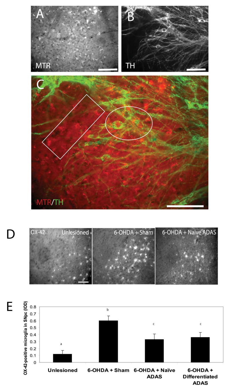
Figure 3. Transplanted ADAS cells engraft in 6-OHDA lesioned brains and attenuate microglial activation.

A, Mitotracker labeled ADAS cells in midbrain sections of rats lesioned with the oxidative neurotoxin 6-hyroxydopamine (see Materials and Methods). B, Expression of the dopaminergic marker TH is not detectable in ADAS cells after transplantation. C, In merged images, the white box indicates a region with singly labeled (MTR-positive) ADAS cells and the white oval indicates a region where MTR-labeled ADAS cells (red) were in close proximity to TH-positive (green) neuron cell bodies. D, Representative images of OX-42-positive microglial staining in SNpc of unlesioned, 6-OHDA-lesioned/sham-transplanted, and 6-OHDA-lesioned naïve ADAS cell-transplanted animals reveal attenuation of neuroinflammatory response in animals that received the ADAS cell transplants compared to lesioned animals that received a sham-transplant. E, Quantification of microglial burden in SNpc, IOD = integrated optical density (see Materials and Methods). Values are means ± S.E.M. Values were compared by one-way ANOVA followed by Tukey-Kramer HSD post-hoc test. Groups with different letters are significantly different at p<0.05. Scale bars=200 um.
