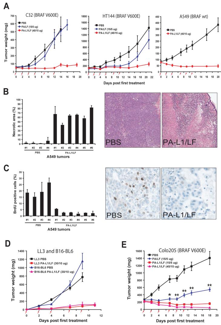
Fig. 2.
PA-L1/LF displays broad and potent anti-tumor activity regardless of the BRAF mutation status of the tumor.
(A) Nude mice bearing human C32 melanoma (left), HT144 melanoma (middle), or A549/ATCC lung carcinoma (right) were injected (i.p.) with 6 doses of PBS, PA/LF, or PA-L1/LF as indicated by red arrows (n=10 for each group). Weights of tumors in this and the following experiments are expressed as mean tumor weight ± s.e.m.
(B) PA-L1/LF causes extensive necrosis of A549/ATCC tumors. A549/ATCC tumor-bearing nude mice were treated with 4 doses of 30/10 μg of PA-L1/LF or PBS (at days 0, 2, 4, and 7). Two hours after injection of BrdU, tumors were dissected and subjected to histological analysis. H&E staining shows extensive toxin-dependent necrosis of a representative tumor treated with PA-L1/LF, which is observed in all the toxin-treated A549/ATCC tumors.
(C) BrdU incorporation assay reveals remarkable DNA synthesis cessation in PA-L1/LF-treated but not PBS-treated A549/ATCC tumors. The tumor sections analyzed in (D-E) were stained with an antibody against BrdU 2 h after systemic administration of BrdU. Note, BrdU positive cells are easily detected in PBS-treated tumors, but hardly detected in viable areas of the toxin-treated tumors.
(D) C57BL mice bearing mouse B16-BL6 melanomas or LL3 Lewis lung carcinomas were treated (i.p.) with 5 doses of PBS or PA-L1/LF as indicated (n=10 for each group).
(E) PA-L1/LF displays much stronger anti-tumor activity than PA/LF. Nude mice bearing Colo205 colon carcinoma were treated (i.p.) with 6 doses of PBA, PA/LF, or PA-L1/LF as indicated (n=10 for each group). A significant difference (*, p<0.05; **, p<0.01) is shown between 15/5 μg of PA-L1/LF and 15/5 μg of PA/LF treated tumors.

