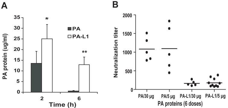
Fig. 3.
Increased plasma half-life and decreased immunogenicity of the MMP-activated protective antigen.
(A) PA-L1 has a longer plasma half-life than PA. Mice were injected (i.v.) with 100 μg of PA or PA-L1, euthanized at 2 h or 6 h, blood samples were collected, and PA protein concentrations were measured using ELISA. There is a significant difference (*, p<0.05; **, p<0.01) between PA and PA-L1. (B) C57BL/6 mice were injected i.p. with 6 doses of 5 or 15 μg of wild-type PA or PA-L1, respectively within a period of two weeks. Ten days later, the mice were bled, and the titers of the serum neutralizing antibodies against PA measured in a cytotoxicity assay using mouse macrophage RAW264.7 cells challenged with LT (75 ng/ml each of PA and LF). The titers of the PA neutralizing antibodies were expressed as mean of fold dilution ± s.e.m of the sera that could protect 50% of RAW264.7 cells from LT treatment. Note that the neutralizing activities from the mice treated with wild-type PA were approximately 6-fold higher that those from PA-L1 treated mice: PA vs. PA-L1 (6×5 μg): 1097 ± 272 vs. 178 ± 36, p=0.0002; PA vs. PA-L1 (6×30 μg): 1081 ± 142 vs. 162 ± 31, p=0.0004.

