Fig. 6.
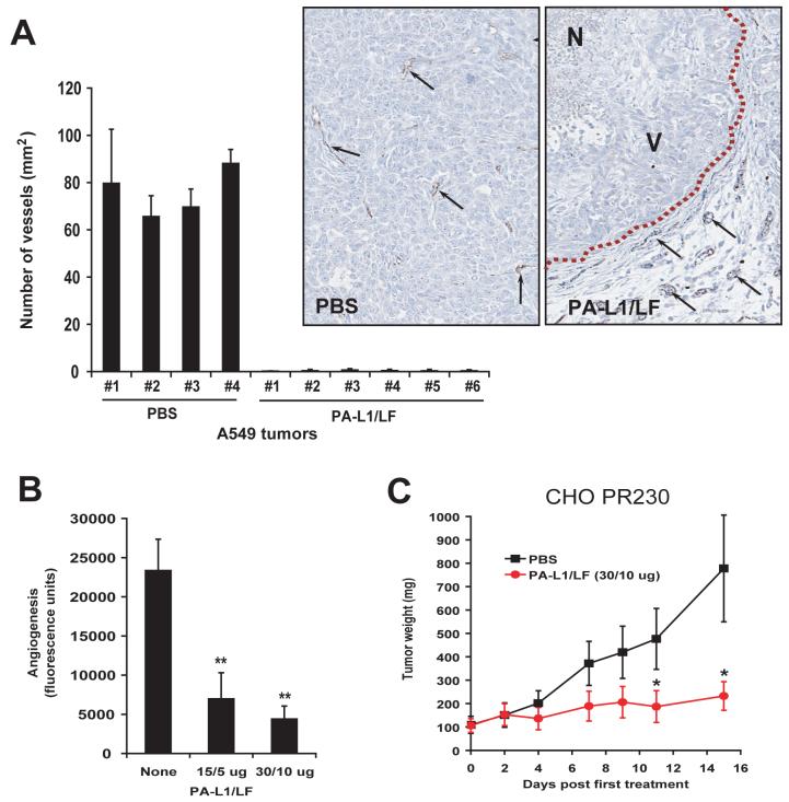
PA-L1/LF demonstrates potent anti-tumor vasculature and angiogenesis activities.
(A) Sections of A549/ATCC tumors treated with PBS or PA-L1/LF, as described inFig. 2Bwere stained with an antibody against the endothelial cell marker CD31. CD31-positive structures were quantified using the Northern Eclipse Image Analysis Software (Empix Imaging, North Tonawanda, NY). In inserts, black arrows point to the examples of CD31 positive endothelial cells; dash line, the boundary between the tumor and its surrounding normal tissues. N, necrotic area; V, area with viable cancer cells.
(B) Directed in vivo angiogenesis analysis demonstrates that PA-L1/LF can inhibit tumor cell independent in vivo angiogenesis. There is a significant difference (**, p<0.01) between the angioreactors treated with PBS (n=8) and treated with PA-L1/LF (15/5 μg, n=8; 30/10 μg, n=10).
(C) Anthrax toxin receptors-deficient CHO tumors are susceptible to PA-L1/LF. CHO PR230 tumor-bearing nude mice were injected (i.p.) with 6 doses of 30/10 μg of PA-L1/LF as indicated (n=6 for each group). There is a significant difference (*, p<0.05) between the tumors treated with PA and PA-L1.

