
Fig. 2.
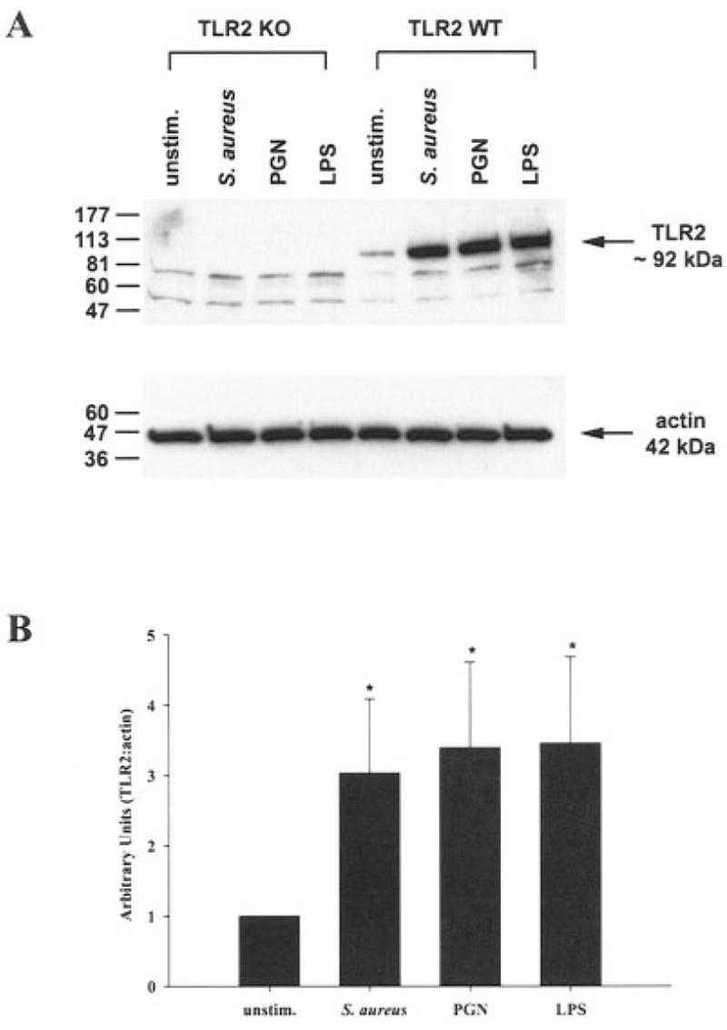
Both S. aureus and peptidoglycan (PGN) enhance TLR2 protein expression in primary microglia. TLR2 knockout (KO) and wild-type (WT) primary microglia were stimulated with either 107 heat-inactivated S. aureus, 10 μg/ml PGN, or 100 ng/ml LPS. Protein extracts from whole cell lysates were prepared 24 h following stimulation and evaluated for TLR2 expression by Western blotting as described in the Materials and Methods. Results are presented as the raw gel data (A) and quantitative analysis of TLR2 expression by densometric scanning (B). For quantitation in B, the pixel intensity of each TLR2 band from WT microglia was normalized to the amount of actin included as a “housekeeping” gene. Results are expressed in arbitrary units as the ratio of TLR2 to actin and represent the mean ± SD of three independent experiments. Significant differences are denoted with asterisks (*P < 0.05).