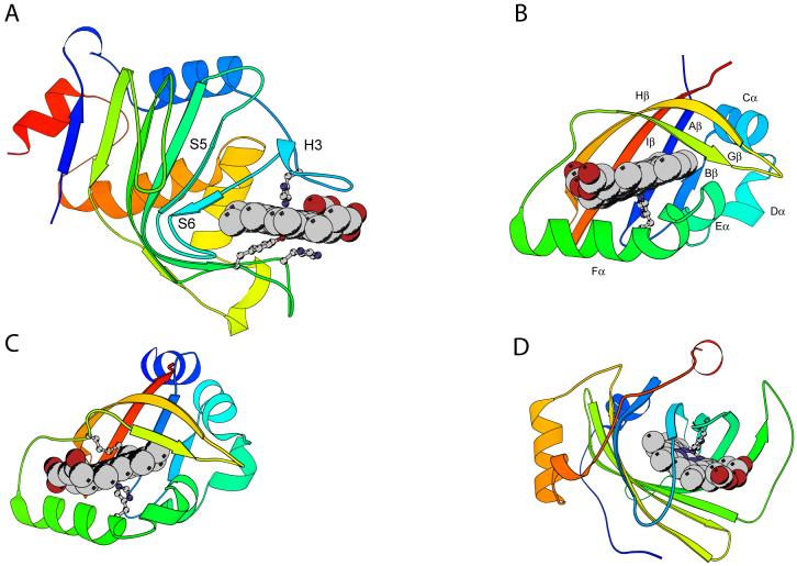
Fig. 3.
Ribbon diagrams of the holoproteins discussed in this review, non α-binding sites. The heme group and iron axial ligands are shown; the N-terminus is blue and the C-terminus is red. Elements of structure mentioned in sections 3 and 4 are marked. A: Serratia marcescens hemophore HasA (1B2V) with axial His32 and Tyr75, assisted by His83; B: heme-containing PAS domain of Bradyrhizobium japonicum FixL (1XJ6; residues 153 to 257), with axial His200 and key hydrophobic residue Ile204; C: Escherichia coli direct oxygen sensor (Dos) heme domain (1S66), with axial His77 and Met95; D: Rhodnius prolixus nitrophorin 2 (1EUO), with axial His57.

