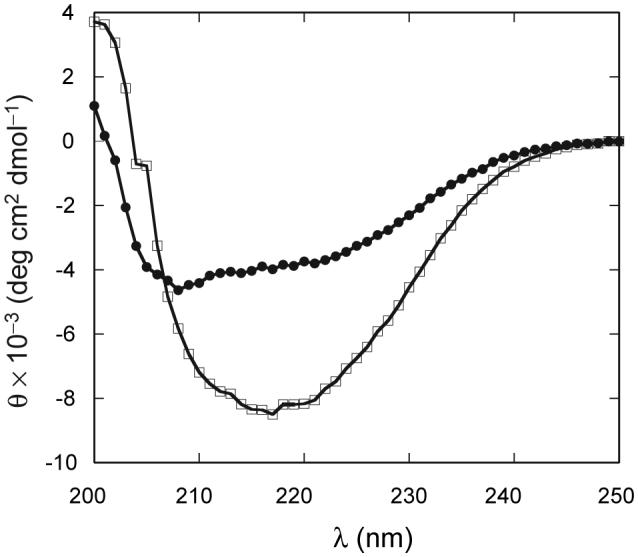
Fig. 7.

Comparison of far UV-CD spectra of apo (●) and met (□) bjFixLH. Sample conditions were pH 9.0 in 20 mM borate 250 mM NaCl at 25 °C. The spectrum of apo bjFixLH was recorded at a protein concentration of ∼10 μM and that of met bjFixLH at ∼25 μM on a heme basis.
