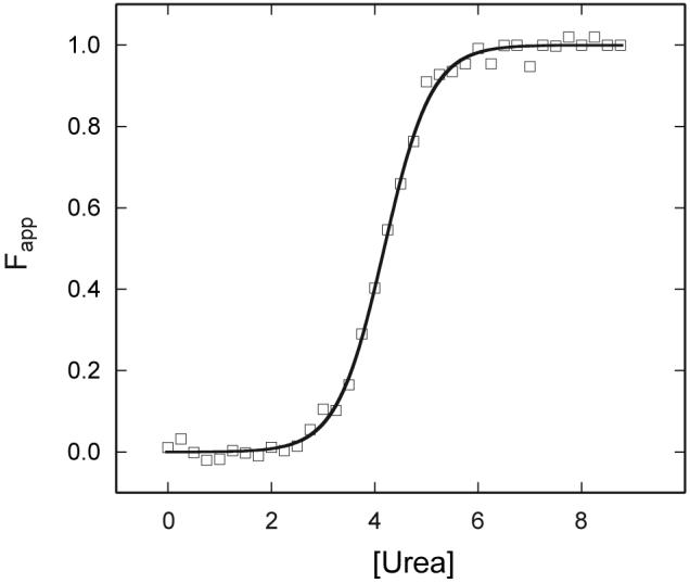
Fig. 9.

Urea denaturation of met bjFixLH as monitored by far UV-CD spectroscopy (222 nm). The apparent fraction of unfolded protein is presented at pH 9.0 (20 mM borate, 250 mM NaCl). Final protein concentration was ∼5 μM. The fit is shown by the line, and one representative set of data is shown by the symbols.
