Figure 3.
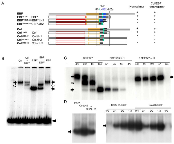
The HLH motif mediating the formation of COE dimers. (A) Diagrammatic representation of full-length mouse EBF/COE1 and Drosophila Col and various carboxy-terminal or/and internal deletions used for Electrophoretic mobility shift assays EMSA (see also Additional file 4). The colour code is as in Fig.1. The deleted helix motif is preceded by Δ in the second column. The homodimer and heterodimer (EBF/Col) columns summarize the data from EMSA;. (B,C,D) EMSA with the 32P labelled mb-1 probe and variants of Col and EBF as indicated above each lane. Homodimers are indicated by black arrows and heterodimers by open arrows, respectively, with the presence of a truncated form of either Col or EBF indicated by*. The free probe is marked by an arrowhead (only shown in B). (B) Col or EBF and EBF* are able to form homo and heterodimers. (C) The H1 α-helix is required for either Col or EBF* to form dimers. (D) The H2 α-helix is also required for Col to form either homodimers or heterodimers with EBF*, left and right lanes, respectively. Two different deletions were tested (see A).

