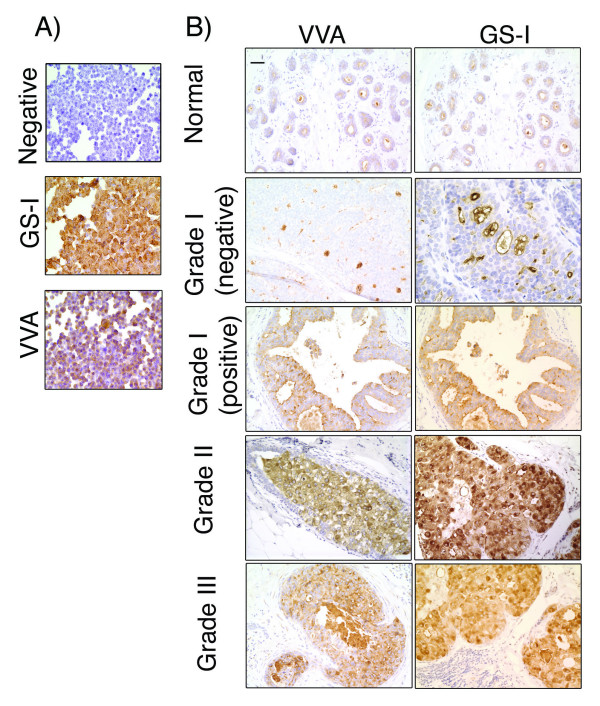
Figure 3.
Lectin histochemical staining of MDA-MB-231 breast cancer cell line and ductal carcinoma in situ.A) Cytospin slides of MDA-MB-231 cells, which were grown in vitro and harvested using enzyme-free buffer. The negative control and staining with GS-I and VVA are shown. MDA-MB-231 breast cancer cell line shows cytoplasmic staining with both lectins and did not show any staining in the absence of the lectins (Negative). 40× Magnification. B) Staining of normal breast tissues and tumor samples of different nuclear grade with VVA and GS-I lectins. 20× magnification, bar equals 50 μm.

