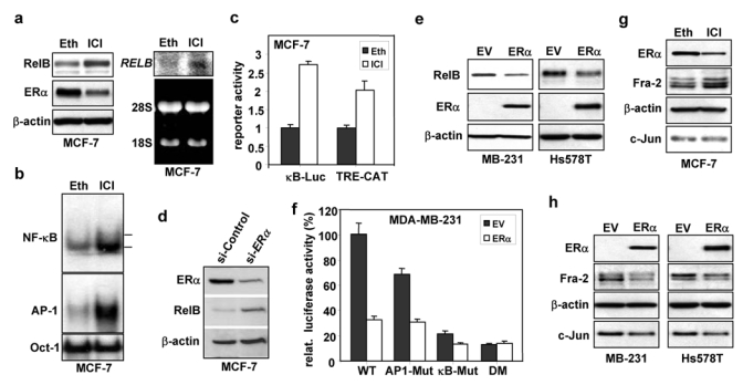
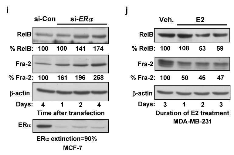
Figure 3. ERα represses NF-κB and AP-1 activities and reduces RelB levels.


(a–b) MCF-7 cells were treated with 1 μM ICI 182,780 (ICI) or vehicle ethanol (Eth) for 48 h. (a) WCEs (25 μg) were analyzed by immunoblotting (left panel). RNA was subjected to Northern blotting and ethidium bromide staining (right panel). (b) Nuclear extracts were subjected to EMSA, as in Fig. 1e. (c) MCF-7 cells were transiently transfected, in triplicate, with 0.5 μg NF-κB (κB-Luc) or AP-1 (TRE-CAT) element driven constructs and 0.5 μg SV40-β-gal. Post-transfection (6 h), cells were treated with ICI 182,780 or ethanol for 40 h. Normalized values are presented as the mean ± S.D. from three experiments (control set to 1). (d) MCF-7 cells were transfected with si-ERα or GFP control siRNA. After 3 successive 48 h transfections, WCEs were analyzed by immunoblotting. (e) Cells, growing in medium depleted of steroids, were transfected with 10 μg ERα expression vector or EV DNA for 6 h and then treated for 40 h with 10 nM E2. WCEs were analyzed by immunoblotting. (f) Cells in steroid-free medium were transfected, in triplicate, with 0.5 μg of the indicated p1.7 RELB promoter-Luc vector, 1 μg ERα expression vector or EV pcDNA3, 0.5 μg SV40-β-gal, and EV DNA (3.0 μg total). After 6 h, cells were treated for 40 h with 10 nM E2. Normalized data are presented relative to the WT reporter with EV DNA (mean ± S.D from three experiments). (g) WCEs from MCF-7 cells, treated as in part a, were analyzed by immunoblotting. (h) MDA-MB-231 and Hs578T cells, transfected with an ERα expression or EV DNA, were processed as in part e. (i) MCF-7 cells, transfected with si-ERα or control siRNA, were lysed after 1, 2 or 4 days and WCEs analysed. Values for RelB and Fra-2 normalized to β-actin relative to control (set at 100%) are presented below. (j) Stable MDA-MB-231 cells expressing ERα, cultivated in medium depleted of steroids, were treated with 10 nM E2 as indicated. Values for RelB and Fra-2 normalized to β-actin in WCEs, relative to control, are given.
