
Figure 4. RelB expression promotes mesenchymal phenotype of breast cancer cells.
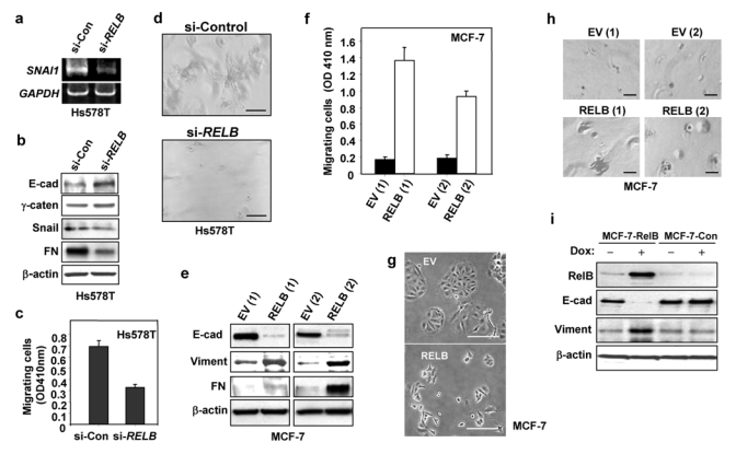
(a–d) Stable Hs578T transfectants expressing either siRNA RELB (Hs578T/si-RELB) or sense RELB (Hs578T/si-Control) (si-Con) were prepared. Expression of si-RELB decreased levels of RELB mRNA and RelB protein in the nucleus, but had no effect on levels of the p65 NF-κB subunit (see Supplementary Information, Figs. S1b1 and S1b2), consistent with findings of Johansen and coworkers33. (a) RNA was analyzed by RT-PCR for levels of SNAI1 and GAPDH RNA, as a control for equal loading. (b) WCEs (50 μg) were analyzed by immunoblotting for E-cadherin, γ-catenin, Snail, fibronectin (FN), and β-actin. The effects of si-RELB on MDA-MB-231 cells were tested and similar data were obtained (see Supplementary Information, Fig. S1c). (c) Cells were subjected, in triplicate, to a migration assay for 4 h. Cells that migrated to the lower side of the filter quantified by spectrometric determination at OD410nm (mean ± S.D). Data presented are from one representative of 3 experiments. (d) Cells were subjected to Matrigel outgrowth analysis. After 5 days, the colonies were photographed (50× magnification). (e–h) Stable MCF-7 clones expressing either RELB or EV DNA were isolated: RELB(1), RELB(2), EV(1), and EV(2). Ectopic RelB expression increased NF-κB reporter activity, and levels of cyclin D1, but failed to alter the levels of p65 (see Supplementary Information, Figs. S2a and S2b). (e) WCEs were subjected to immunoblot analysis for E-cadherin (E-cad), fibronectin (FN), vimentin (Viment), and β-actin. While all of the clones showed an increase in fibronectin, the extent of the increase varied. (f) Clones were subjected to a migration assay, as in part (c) (mean ± S.D from three separate experiments). (g) MCF-7 stable clones (1×10⁴ cells ml−1) were plated and after 2 days photographed (50× magnification). (h) Clones were subjected to Matrigel outgrowth analysis. After 15 days of growth, the colonies were photographed (50× magnification). (i) Stable MCF-7 cells with an inducible RelB expression vector were treated with 1 μg/ml doxycyline. After 48 h, WCEs were subjected to immunoblot analysis for RelB, E-cadherin (E-cad), vimentin and β-actin, as above. Scale bar, 100 μm in parts d, g, and h. An uncropped scan of the E-cadherin panel in i is shown in the Supplementary Information, Fig. S5c.