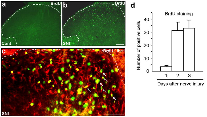
Figure 1. (a–d). Spared nerve injury (SNI) induces proliferation of microglial cells in the spinal cord.

(a) Sham control (Cont) animals show almost no BrdU immunostaining. (b) Three days following SNI, there is a profound proliferation in the ipsilateral dorsal horn. Scale, 100 μm. (c) Double immunostaining of BrdU with the microglial marker Iba1 in the dorsal horn. Note that most BrdU positive cells also express Iba1 (indicated with arrows). White lines show the borders of the dorsal horn. Scale, 100 μm. (d) Number of BrdU-positive cells (per 30 μm-thick section) in the spinal cord dorsal horn. Note that proliferation starts rapidly after SNI.
