
Figure 3. (a–e). Activation of p38 and ERK in the spinal cord after SNI is required for neuropathic pain development.
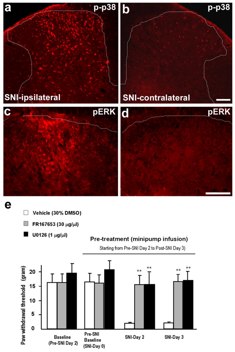
(a–d) Immunohistochemistry shows an increase in phosphorylation of p38 (p-p38, a, b) and ERK (pERK, c, d) in the medial dorsal horn on the ipsilateral side (L4) 3 days after SNI. White lines indicate the borders of the dorsal horn. Scales, 100 μm. (e) SNI-induced mechanical allodynia is prevented by the p38 inhibitor FR167653 or the MEK (ERK kinase) inhibitor U0126. FR167653 (30 μg/μl) or U0126 (1 μg/μl) was infused into intrathecal space via an osmotic pump (0.5 μl/h for 5 days) starting 2 days before SNI. Note that the basal mechanical sensitivity does not change after FR167653 or U0126 infusion. **, P < 0.01, t test, compared to corresponding vehicle controls (30% DMSO), n = 4. Modified from Wen et al., 2007.