
Figure 6. (a–c). Schematic representation of microglial control of pain.
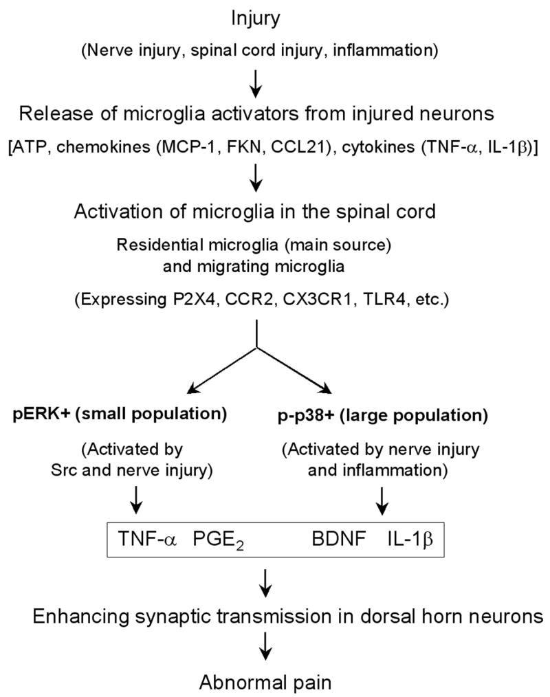
After various injury conditions (such as nerve injury, spinal cord injury, and inflammation), injured or affected neurons (e.g., DRG or spinal cord neurons) can release factors that are capable of activating microglia in the spinal cord. These factors include ATP, chemokines such as MCP-1, fractalkine (FKN), and CCL21, and proinflammatory cytokines such as TNF-α and IL-1β. These microglia activators can bind their receptors on microglia, leading to the activation of microglia. These activated spinal microglia are mainly residential microglia, but may also be migrating microglia from circulation. Activated microglia contain three subpopulations in the spinal cord: pERK + population, p-p38 + population, and unknown population. While ERK is activated by Src and nerve injury, p38 is activated by both nerve injury and inflammation. ERK activation releases TNF-α via activation of TNF-α converting enzyme, whereas p38 activation enhances IL-1β release. Activation of p38 and ERK can also regulate the synthesis of the inflammatory mediators via transcription factor (e.g., NF-κB). Microglia also produce the growth factor BDNF. Finally, these pain mediators collaborate to induce and maintain abnormal pain in injury conditions by enhancing synaptic strength and central sensitization in dorsal horn neurons.