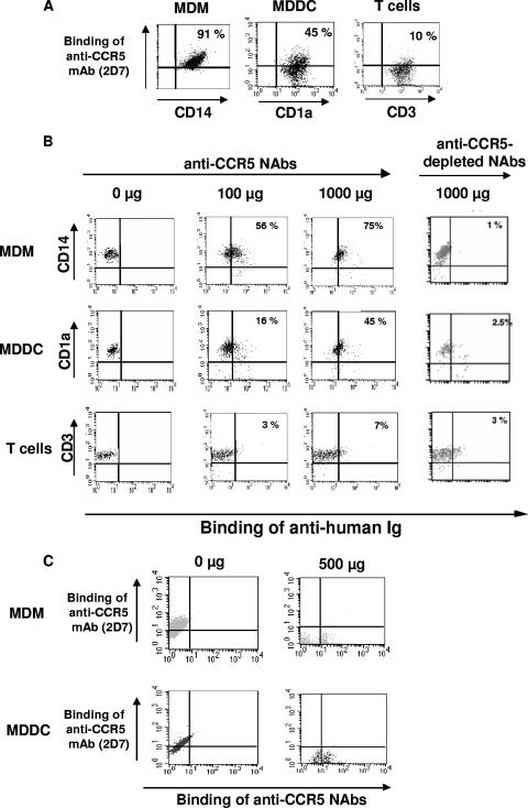
FIG. 5.
Binding of anti-CCR5 NAbs to macrophages (CD14+), immature dendritic cells (CD1a+), and T lymphocytes (CD3+) at the single-cell level. (A) MDMs, MDDCs, and T lymphocytes (105) were incubated with anti-CD14-FITC, anti-CD1a-FITC, and anti-CD3-FITC MAbs, respectively, and with an anti-CCR5 PE MAb (2D5 clone; 10 μg/ml) at 4°C for 30 min. (B) MDMs, MDDCs, and T lymphocytes (105) were incubated with mouse anti-CD14 PE, anti-CD1a PE, and anti-CD3-PE MAbs, respectively, and with either 0, 100, or 1,000 μg/ml of purified anti-CCR5 NAbs or 1,000 μg/ml of anti-CCR5-depleted NAbs at 4°C for 30 min. The cells were subsequently incubated with FITC-conjugated goat anti-human F(ab′)2 antibody at 4°C for 30 min. (C) MDMs and MDDCs (105) were incubated with or without 1,000 μg/ml of purified anti-CCR5 NAbs at 4°C for 30 min. The cells were subsequently incubated with FITC-conjugated goat anti-human F(ab′)2 antibody and an anti-CCR5 PE MAb at 4°C for 30 min. The percentage of positive cells is indicated in quadrants defined according to the relevant isotypic control. The results of one representative experiment of four independent experiments conducted are shown.

