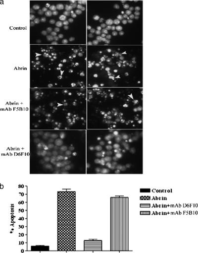
FIG. 8.
(a) Jurkat cells were cultured with abrin (10 ng/ml) in the presence or absence of antibodies (25 μg/ml) for a period of 12 h, stained with acridine orange-ethidium bromide, and analyzed by fluorescence microscopy. Arrowheads point to apoptotic nuclei. (b) In another experiment, the cells were incubated with abrin, fixed in 70% ethanol, and stained with ethidium bromide. The percentage of apoptosis was quantified by using FACScan. The experiment was carried out at least three times. Representative data are presented.

