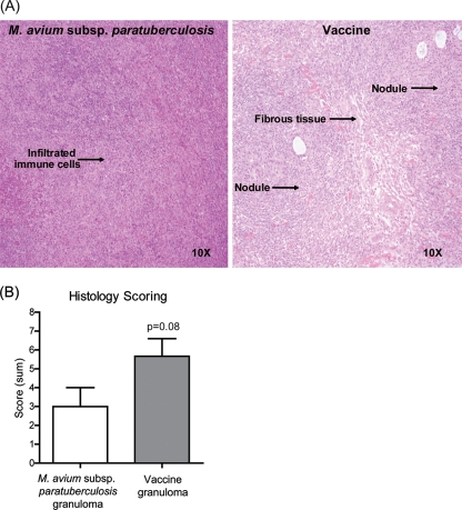
FIG. 1.
Histology of granulomatous lesions induced by M. avium subsp. paratuberculosis or vaccine at 7 days postinoculation. (A) H&E-stained sections of formalin-fixed and paraffin-embedded tissues. Images representative of results from two M. avium subsp. paratuberculosis-inoculated animals and four vaccinated animals are shown. Original magnification is shown. (B) Lesions were scored from 0 to 3 based on the following individual parameters: mature fibrous connective tissue delineation, macrophage infiltration, lymphocyte organization, multinucleated giant cells, necrosis, and mineralization. Individual parameters were summated, and a single collective score was assigned for each lesion which reflects the histological organization of individual lesions. Pooled data from two M. avium subsp. paratuberculosis-inoculated animals and four vaccinated animals are shown.

