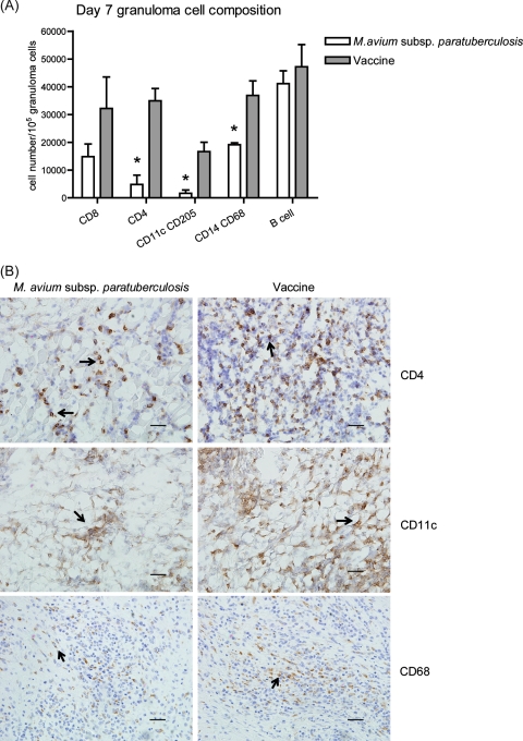
FIG. 2.
Cellular composition of granulomas at 7 days postinoculation. (A) Total cells were recovered from granuloma digests. Phenotype was determined by using immunofluorescent staining for surface markers and flow cytometry. Data are the means ± standard errors of the means of the number of cells expressing a surface marker from two M. avium subsp. paratuberculosis-inoculated animals and four vaccinated animals. *, P < 0.05. (B) Granulomatous lesions were immunohistochemically stained for CD4, CD11c (from frozen sections), and CD68 (from formalin-fixed sections). Arrows indicate positive staining for the specific markers. Images representative of results from two M. avium subsp. paratuberculosis-inoculated animals and four vaccinated animals are shown. Bars represent 50 μm.

