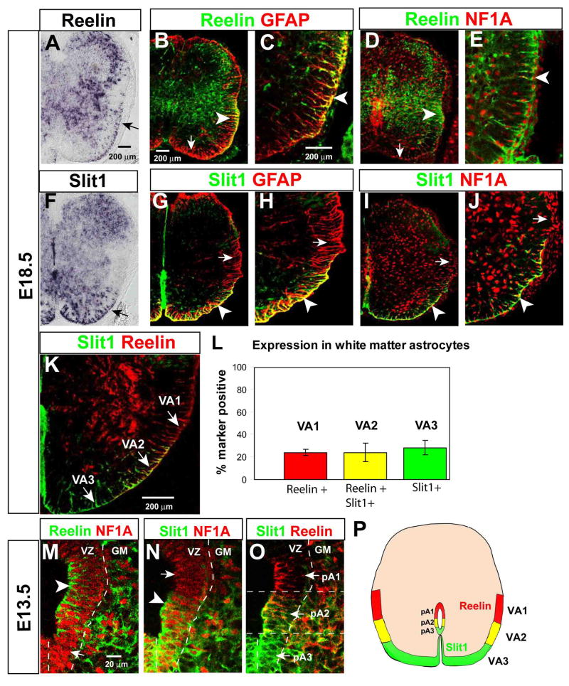
Figure 1. Reelin and Slit1 define astrocyte subpopulations in the ventral spinal cord.
(A, F) In situ hybridization for mouse Reelin and Slit1 mRNAs. Arrows indicate expression in ventral white matter. (B–E) Double-label immunohistochemistry for Reelin and GFAP (B–C) or NFIA (D–E). Arrowheads indicate double-positive cells in lateral white matter, arrows Reelin− astrocytes in ventral white matter. (G–J) Double-label immunohistochemistry for Slit1-GFP and either GFAP (G–H) or NFIA (I–J), in Slit1GFP/+ embryos. Arrowheads indicate Slit1+ astrocytes in ventral white matter, arrows Slit1− astrocytes in the lateral white matter. (K) Double labeling for Reelin and Slit1-GFP reveals three subpopulations of ventral astrocytes (arrows). (L) Quantification of the percentage of NFIA+ astrocytes expressing each of the three markers. Data represent the mean±S.E.M. of 5–6 sections/embryo from 3 embryos. (M–O) Triple labeling for Reelin, Slit1-GFP and NFIA in the E13.5 ventral VZ, displayed as pairwise comparisons from the same section. Arrowheads and arrows in (M, N) indicate NFIA+ cells that are positive or negative, respectively, for Reelin (M) or Slit1 (N). (O) Overlay of Slit1 and Reelin reveals three adjacent progenitor domains in the VZ. (P) Composite schematic illustrating positions of VA1–VA3 astrocytes in the white matter at E18.5, and corresponding progenitor domains (pA1–3) in the VZ at E13.5.

