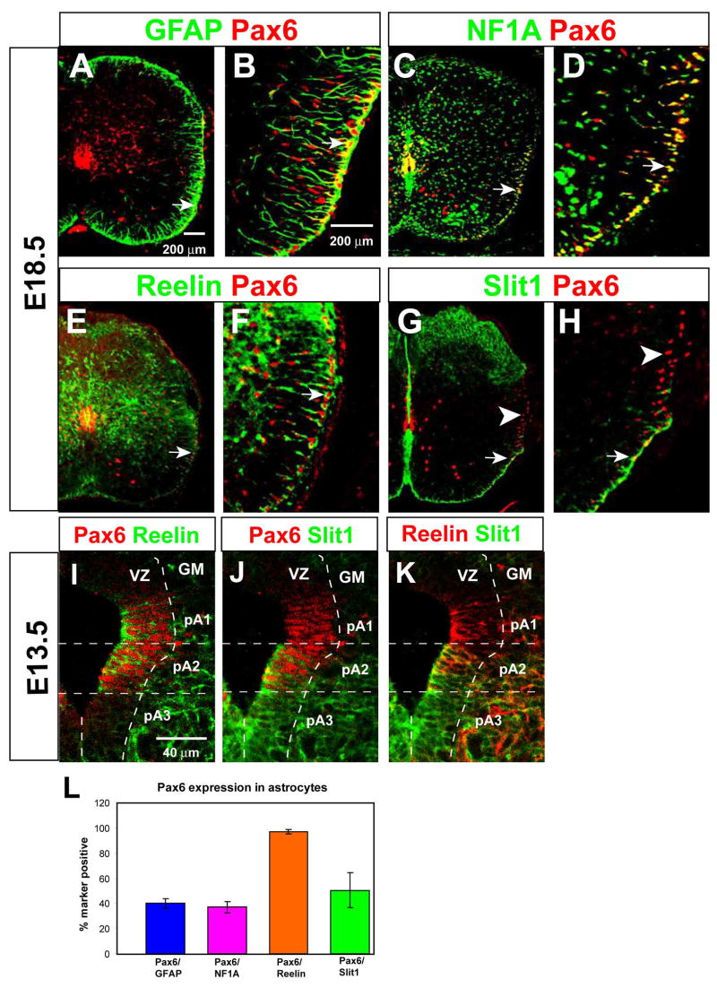
Figure 2. Expression of Pax6 in Reelin+ astrocytes and their precursors.
(A–H) Double-labeling for Pax6 (red) and the indicated markers (green) at E18.5. (B, D, F, and H) are higher magnification views of the areas indicated by arrows in (A, C, E and G), respectively. (I–K) Triple labeling for Pax6, Reelin, and Slit1-GFP in the E13.5 ventral VZ, displayed as pairwise comparisons from the same section. (L) Quantification of Pax6 expression among total (GFAP+ or NFIA+) astrocytes, and Reelin+ or Slit1+ WMAs.

