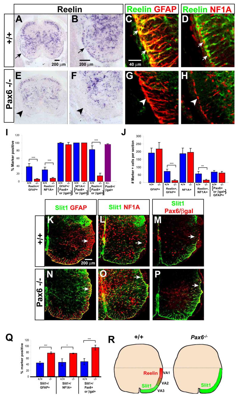
Figure 3. Astrocyte subtype conversion in Pax6−/− mice.

(A–B, E–F) In situ hybridization for Reelin mRNA in E18.5 spinal cord of wild-type (A–B) and Pax6 mutant (E–F) embryos. (B) and (F) are higher-magnification views of (A) and (E), respectively. Arrows in (A, B) indicate Reelin+ cells in the white matter. (C–D, G–H) Double-labeling for Reelin and GFAP (C, G) or NFIA (D, H) in wild-type (C–D) and Pax6 mutant (G–H) embryos. (I–J) Quantification of Reelin expression by WMAs in E18.5 wild type (blue bars) and Pax6−/− (red bars) spinal cord. . (I) The percentage of total GFAP+ or NFIA+ WMAs expressing Reelin is significantly reduced in the Pax6lacZ/lacZ mutant (***, p<.001). (J) The absolute number of GFAP+ and NFIA+ astrocytes in the white matter is not changed in the Pax6lacZ/lacZ spinal cord, nor is the average number of Pax6+ GFAP+ astrocytes. (*** and **, p<.001 and .002, respectively). . The data are derived from 5 wild type and 6 mutant embryos from 3 independent litters. (K–P) Spinal cord sections from Pax6+/+; Slit1GFP/+ (K–M) and Pax6lacZ/lacZ; Slit1GFP/+ (N–P) embryos double-labeled for the indicated markers. Arrows indicate VA1 astrocytes (K–M) that exhibit de-repression of Slit1 in the mutant (N–P). Red staining in (M, P) represents anti-Pax6 (M) or anti-βgal (P). (Q) Slit1 expression among total astrocytes (GFAP+ or NFIA+) is significantly increased in Pax6−/− embryos (*** and *, p = .0003, and .012, respectively). (H) Schematic illustrating the changes in Reelin and Slit1 expression by ventral astrocytes in the Pax6 mutant.
