
Figure 5. Nkx6.1 is co-expressed by Slit1+ astrocytes and their precursors.
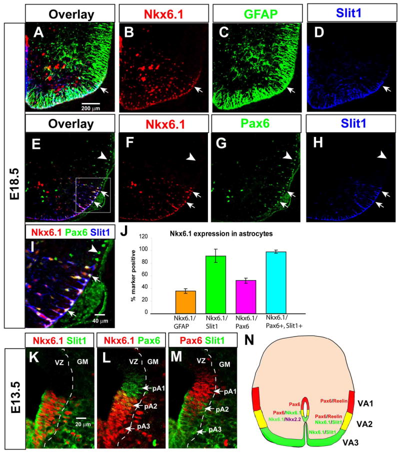
(A–I) Triple-labeling for Nkx6.1, GFAP and Slit1-GFP (A–D) or Nkx6.1, Pax6 and Slit1-GFP (E–I). (I) is a higher power image of the boxed area in (E). Note that all Slit1+ cells in this domain (boxed area, E) are Nkx6.1+ and Pax6+ (E–G, I, arrows, white nuclei), while all Pax6+ cells dorsal to the boundary of Slit1 expression are Nkx6.1− (E–G, I, arrowheads). (J) Quantification of Nkx6.1 expression in astrocyte sub-populations. Nkx6.1 is expressed in > 90% of Slit1+ astrocytes (green bar). (K–M) Triple antibody labeling for Nkx6.1, Slit1-GFP and Pax6 in the E13.5 VZ, displayed as pairwise comparisons from the same section. The three progenitor domains are indicated (L, M, arrows). “GM,” gray matter. (N) Composite schematic illustrating relationship between the domains of Reelin and Slit1 expression, and those of Pax6 and Nkx6.1 expression, in the white matter at E18.5 and in the VZ at E13.5.