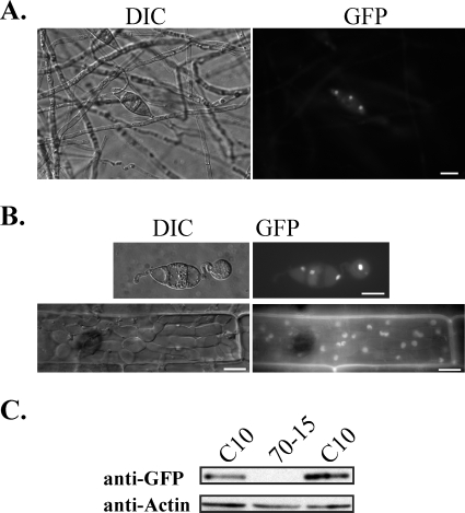
FIG. 6.
Expression and localization of Mig1-GFP in transformant C10. (A) GFP signals were observed in the nuclei of conidia but not in aerial hyphae or conidiophores harvested from 9-day-old oatmeal cultures. The same field was examined under differential interference contrast (DIC) and epifluorescence microscopy (GFP). Bar = 10 μm. (B) GFP signals were preferentially localized to the nuclei in appressoria formed on glass coverslips and infectious hyphae produced in rice leaf sheath epidermal cells. Bars = 10 μm. (C) Western blot analysis with proteins isolated from vegetative hyphae of the wild-type strain 70-15 and transformant C10. An 86-kDa band of the expected size of Mig1-GFP fusion was detected with an anti-GFP antibody.

