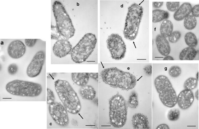
FIG. 2.
Localization of PGA shown by IEM of wild-type and mutant pga strains. Cultures were grown for 24 h at 26°C under standing (b) or shaking (a, c, d, e, f, and g) conditions. Samples were prepared, and PGA was detected with a primary anti-PGA murine MAb and a secondary, gold-labeled anti-murine Ig antiserum (Materials and Methods). Sample identities: MG1655 (a); TRMG1655 (csrA::kanR) (b and c); TRMG1655 ΔpgaA (d); TRMG1655 ΔpgaB (e); TRMG1655 ΔpgaC (f); and TRMG1655 pgaD::cam (g). Note the gold labeling around the perimeter and between cells of the wild-type, standing pga strain culture (b) and the decreased labeling of cells from the shaken culture (c), with label retention at the cell poles (arrows). Polar localization of PGA was observed in 76% (125/165) of longitudinally sectioned cells from 17 different fields. In contrast, the ΔpgaA and ΔpgaB strains from the shaken cultures retained strong circumferential labeling with shaking (d and e). Labeling patterns of ΔpgaA and ΔpgaB mutants under standing conditions (not shown) were essentially identical to those observed under shaking conditions. Size bars are 400 nm in length.

