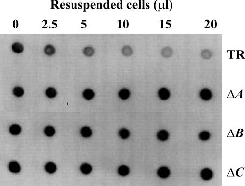
FIG. 3.
Adsorption of anti-PGA MAb to test for surface-exposed PGA on ΔpgaA and ΔpgaB mutant cells. Equal quantities of cells of TRMG1655 (TR) and its isogenic pga mutants were harvested, resuspended, and incubated at several concentrations with the anti-PGA MAb for 1 h. Antibody remaining in the supernatant solution after removal of cells was detected by immunoblotting (Materials and Methods). The reactions across the top and bottom rows depict the results for positive and negative experimental controls, respectively, for surface-exposed PGA. ΔA, ΔpgaA; ΔB, ΔpgaB; ΔC, ΔpgaC.

