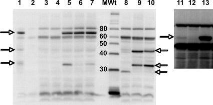
FIG. 6.
Western blots of wild-type and mutant PgaB proteins. Cultures were grown, harvested, and analyzed for PgaB protein by Western blotting using anti-HmsF antibody (Materials and Methods). Strain identities were as follows: 1, Y. pestis KIM6+; 2, Y. pestis KIM6; 3, TRMG; 4, TRMG ΔpgaB(pCR2.1 TOPO); 5, TRMG ΔpgaB(pCRpgaB); 6, TRMG ΔpgaB(pD115A); 7, TRMG ΔpgaB(pH184A); 8, TRMG ΔpgaB(pCRpgaB271); 9, TRMG ΔpgaB(pCRpgaB410); 10, TRMG ΔpgaB(pCRpgaB516). MWt indicates the molecular mass standards and their corresponding sizes in kDa. The horizontal arrows mark the positions of PgaB (HmsF) full-length and partial polypeptides that were detected by the antiserum. Lanes 1 to 10 depict samples separated on a 12.5% gel. Lanes 11 to 13 depict the same samples as in lanes 3 to 5 separated on a 7.5% gel and overexposed to reveal the PgaB native protein (∼74 kDa) from TRMG1655. The Y. pestis and E. coli proteins were loaded at 5 μg and 50 μg per lane, respectively.

