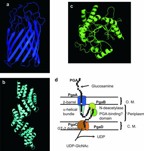
FIG. 7.
HHpred predictions of Pga protein domain structures (a to c) and a hypothetical model for Pga protein assembly in the cell envelope (d). Structural predictions were derived by a threading algorithm using the best domain matches for PgaA C-terminal domain (FadL protein of E. coli) (a), PgaA N-terminal domain (human nucleoporin O-linked GlcNAc transferase) (b), and PgaB C-terminal domain (LacZ of T. thermophilus) (c). The amino acid sequences of these domains are 518 to 807, 65 to 515, and 333 to 646, respectively. The N-terminal domain of PgaB and the sole domain of PgaC belong to carbohydrate esterase (N-deacetylase) family 4 and GT-2 glycosyltransferase family, respectively (50). No prediction was obtained for PgaD. (d) The model for PGA synthesis and Pga protein localization in the cell envelope is discussed in the text. O.M., outer membrane; C.M., cytoplasmic membrane.

