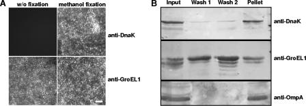
FIG. 2.
Surface localization of C. pneumoniae GroEL1. (A) Chlamydial GroEL1 protein could be detected on Percoll gradient-purified EBs without (w/o) fixation as well as after methanol fixation using indirect immunofluorescence microscopy. Antibodies directed against the intrachlamydial DnaK protein could detect the antigen only after methanol fixation. Bar, 1 μm. (B) Percoll gradient-purified EBs were washed twice with PBS; the input, wash fractions 1 and 2, and pellet were separated by SDS-PAGE and analyzed by Western blot analysis using antibodies against chlamydial OmpA, DnaK, and GroEL1. The wash fractions loaded contained three times as much protein as the pellet samples. Data are representative of several separate experiments.

