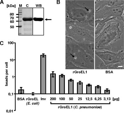
FIG. 3.
Adhesion of rGroEL1-coated latex beads to HEp-2 cells. (A) Coomassie blue-stained SDS-PAGE of the affinity-purified rGroEL1 protein after expression in E. coli (lane C) and analysis of purified rGroEL1 by Western blot analysis using an antibody against the N-terminal His tag (lane WB; the arrow marks the rGroEL1 protein band). Lane M, molecular mass markers. (B) Latex beads were coated with BSA or His-tagged recombinant proteins as indicated (protein concentration during coating, 200 μg/ml). Coated beads were incubated with HEp-2 cells. Adhesion of protein-coated latex beads was visualized by phase-contrast microscopy (magnification, ×63). Arrows indicate latex beads attached to HEp-2 cells. Bar, 10 μm. (C) Dose-dependent adhesion of rGroEL1-coated beads to HEp-2 cells. Beads were coated with different rGroEL1 protein concentrations. The control beads (BSA, Inv, and E. coli GroEL) were always coated with a protein concentration of 200 μg/ml. Results of the adhesion experiments are given as bound beads per HEp-2 cell (n = 1,000 HEp-2 cells; number of experiments = 4). Data shown are means ± standard deviations.

