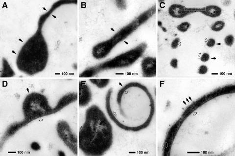
FIG. 3.
Transmission electron micrographs of ultrathin sections of morphological variants of protrusions from strain SSD-17BT. (A) Central body with one “tentacle-like” protrusion (relaxed state); the surface of the cell is coated by a diffuse electron-dense layer (arrows). (B) Detail of “tentacle-like” protrusions (relaxed state); the surface of the cell is coated at both sides by a diffuse electron-dense layer (arrows). (C) Longitudinal section of a helical protrusion (contracted stage); several windings are cross sectioned. (D to F) Cross sections of helical protrusions. The outer side of the rings (C), lateral protuberances (D), and helixes (E and F) are “rough” (black arrows) compared to the inner surface (open arrows). The cytoplasmic membrane can be recognized locally (C, framed area; F, circle).

