FIG. 1.
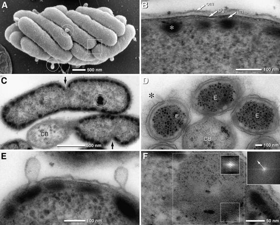
SEM and TEM micrographs of “Chlorochromatium aggregatum” and the epibionts, “Chlorobium chlorochromatii” cells, in the symbiotic state and in pure culture. (A) SEM of “Chlorochromatium aggregatum” showing the epibionts tightly packed; the cell surface appears rough and exhibits numerous thin filaments interconnecting neighboring cells (frame). Epibionts exhibit numerous bulb-shaped protrusions (circles). (B) TEM detail of the epibiont cell with typical gram-negative envelope (om, outer membrane; pg, peptidoglycan; im, inner membrane) and chlorosomes attached to the cytoplasmic membrane (asterisk). (C) TEM of epibionts (E)—attached to a central bacterium (CB)—revealing asymmetric cell division (arrows). (D) TEM of consortia after cryofixation by high-pressure freezing showing with high contrast the hair-like filaments (asterisk) and the undulating outer membrane (the chlorosomes appear electron translucent due to extraction during freeze substitution) of the epibionts (E, epibiont; CB, central bacterium). (E) TEM of epibiont bulb-shaped protrusions which are formed from the outer membrane by local enlargement of the periplasmic space. (F) Lipid body-like globule of pure culture of “Chlorobium chlorochromatii” exhibiting a myelin-like structure which is enhanced after autocorrelation (small insets) with a spacing of 3.5 nm, determined by FFT (upper right inset; the arrow indicates the first-order reflex) of the dotted area.

