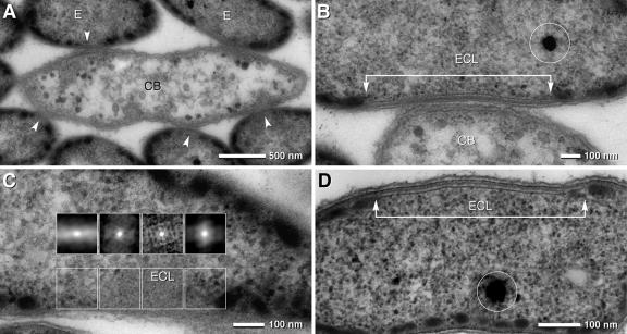
FIG. 2.
TEM micrographs of consortia and epibionts in the symbiotic state and in pure culture. (A) TEM of a longitudinal ultrathin section of a consortium with a central bacterium (CB) with the shape of an elongated ellipse (with the beginnings of cell division) and several attached epibionts (E); at the site of attachment, the cytoplasmic membrane of the epibionts is free of chlorosomes (arrowheads). (B) Area of attachment of the epibiont cell and the central bacterium (ECL, epibiont contact layer) which is characterized by a laminar layer (arrow bar) and the absence of chlorosomes; the osmiophilic globule (circle) represents polyphosphate. (C) Autocorrelation of an oblique section of an ECL revealing a pattern of regularly arranged particles (insets); at transition zones from the ECL to the cytoplasm, the pattern disappears (upper insets, outer left and right). (D) TEM of an ECL of “Chlorobium chlorochromatii” in pure culture; although the ECLs are present only in approximately 20% of the cells, their architecture is identical to that formed in symbiosis; the osmiophilic globule (circle) represents polyphosphate.

