FIG. 3.
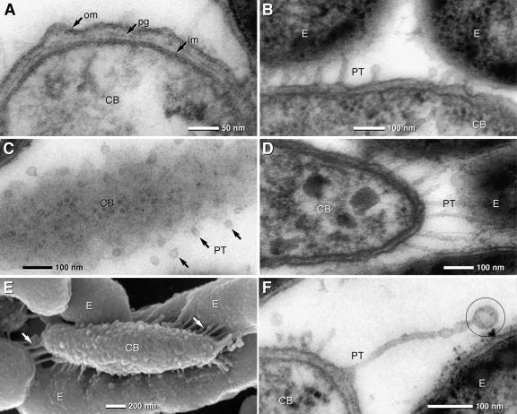
TEM of ultrathin sections and SEM micrographs of the central bacterium (CB) and epibionts (E) of “C. aggregatum.” (A) TEM detail of the central bacterium cell with a typical gram-negative envelope (om, outer membrane; pg, peptidoglycan; im, inner membrane). (B and C) TEM of PT formed by the outer membrane of the central bacterium which are in contact with the outer membrane of the epibionts (B). In tangential sections of the central bacterium, PT are visible as small circular membrane cross sections with diameters in the range of 25 nm (C, arrows). (D and E) The lengths of the PT depend on the distance between the central bacterium and the epibionts. PT are rather straight and appear under tension when in contact with the epibionts, which is best seen in 150-nm sections (D) and SEM micrographs of a partially desegregated consortium (E). The central bacterium exhibits a knobby surface, with numerous PT (arrows) attached to the epibionts (E). (F) Unattached PT show terminal vesiculation (circle).

