FIG. 4.
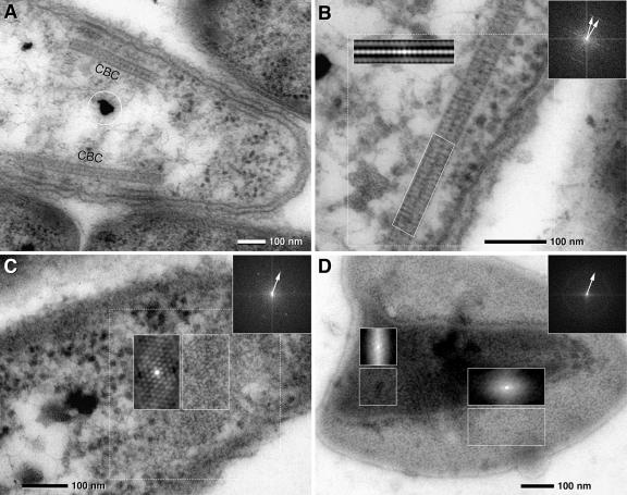
TEM micrographs of ultrathin sections of the central bacterium of “C. aggregatum” revealing paracrystalline structures (CBC, central bacterium crystal) in situ and after isolation and negative staining. FFTs of dotted areas are presented as insets (upper right; arrows indicate first-order reflexes); autocorrelation is presented in the same size as the analyzed area. (A) Longitudinal section of a central bacterium with two cross-sectioned CBCs located parallel to the cytoplasmic membrane (circle, osmiophilic granules). (B) Detail of a CBC showing a paracrystalline zipper-like organization in the cross sections; two less-electron-dense layers are bordered and separated by electron-dense layers. Autocorrelation and FFT of the CBC reveal a 9-nm periodicity with the orientation of the subunits either vertical or oblique to the axis (two 9-nm FFT reflexes). (C) FFT and autocorrelation of a tangential section of the CBC shows a hexagonal pattern of subunits which are 9 nm in diameter. (D) Negative staining of the isolated CBC. Isolated CBCs are frequently observed as double or multiple layers. FFT of the whole image shows a Debye-Scherrer ring corresponding to a distance of 9 nm, indicating a lower degree of subunit orientation of the CBC than that found in situ; autocorrelation shows local patterns of subunit orientation similar to that of CBC in situ in a tangential ultrathin section (for comparison, see panel C).

