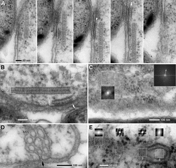
FIG. 5.
TEM micrographs of ultrathin sections of the central bacterium of “C. aggregatum” revealing the CML and MW. (A) Serial sections show that the CML is only visible in two to three consecutive sections, proving that it is locally restricted. The CMLs are found with different thicknesses, suggesting the composition of a monolayer (arrowhead) or bilayer (arrow). (B) The electron density of the outer leaflet of the cytoplasmic membrane (im) is higher at the site of attachment of the CML (arrow). (C) FFT and autocorrelation of a tangential section of the CML shows a hexagonal pattern of subunits which are 9 nm in diameter (the arrow indicates the first-order reflex). (D) Details of the central bacterium showing MW which are formed by invaginations of the cytoplasmic membrane (arrow). (E) Autocorrelation of different areas of a section through a CBC in contact with a MW: an oblique section of the CBC is characterized by striations (1) and tangential sections with hexagonal patterns (2, 3); no significant pattern is visible at the area of the MW (4).

