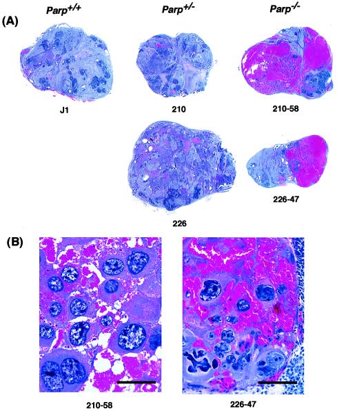
Figure 2.
Photomicrograph of the intratumoral STGCs and hemorrhage in tumors derived from Parp−/− clones. (A) The loupe findings of paraffin sections. Magnification: ×3.2. The hemorrhagic areas, seen as blood lakes, were present only in tumors derived from Parp−/− clones. (B) High-power magnification of the STGCs containing single or multiple megalo-nuclei and eosinophilic cytoplasm. The STGCs are present within the hemorrhagic area. Bars indicate 100 μm. Magnification: ×120.

