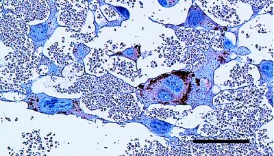
Figure 4.
The STGCs stained with anti-mouse prolactin antibody. The cytoplasmic fractions of STGCs in tumors derived from Parp−/− clone were stained. Some granules in the STGCs are strongly stained. Bar indicates 100 μm. Magnification: ×160. The negative control sections, from which the primary antibody was omitted, showed no positive staining (data not shown).

