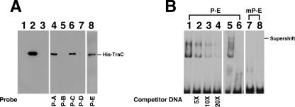
FIG. 5.
Binding of TraC to pSW100. (A) Purified histidine-tagged TraC (lane 2) was added to a binding mixture that contained a biotinylated DNA probe, P-A (lane 4), P-B (lane 5), P-C (lane 6), P-D (lane 7), or P-E (lane 8). A DNA-protein complex was then captured using streptavidin-coated magnetic beads. Proteins bound to the beads were separated by SDS-PAGE and detected by immunoblotting using an anti-histidine tag antibody. In a negative control, a P-A probe was incubated with protein extract from E. coli BL21(DE3)(pET-30b) (lane 1). Lane 3 was loaded with the P-A probe. (B) The P-E probe (lanes 1 to 6) and a mutant P-E probe (mP-E) (lanes 7 and 8) were used to analyze the binding of His-TraC to the P-E region by EMSA. Anti-His antibody was utilized to demonstrate the supershifting of the probe (lane 5). Lanes 6 and 7 were loaded with the P-E and mP-E probes, respectively. Each binding reaction involved 5 ng of biotinylated probe, 6 μg of TraC, and 1 μg of poly(dI-dC). Unlabeled P-E DNA was used to compete the binding (lanes 2 to 4). The protein-DNA complex was separated with a 7% polyacrylamide gel and detected using a LightShift chemiluminescence EMSA kit (Pierce).

