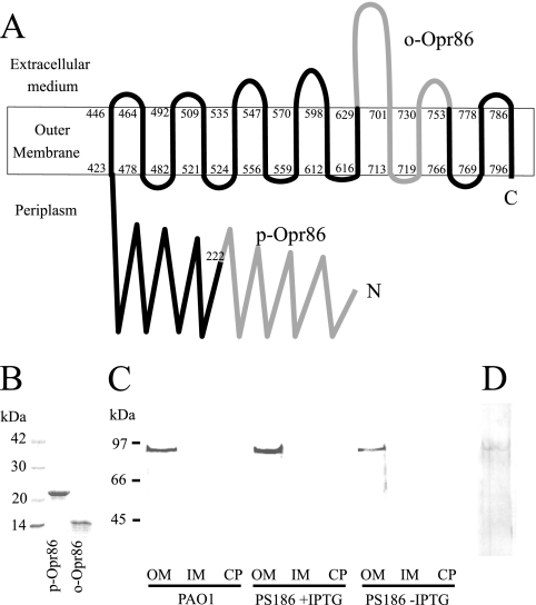
FIG. 4.
(A) Topology prediction of Opr86 in P. aeruginosa. It is predicted that Opr86 has 14 transmembrane domains. The first and last amino acids of each β-strand are indicated. The domains of p-Omp86 and o-Opr86 are shown in gray. The amino (N) and carboxyl (C) termini of the protein are indicated. (B) SDS-PAGE analysis of p-Opr86 and o-Opr86 purification, performed on a 15% gel. Two micrograms of purified protein was applied per well, and the gel was stained with Coomassie brilliant blue. (C, D) Immunoblot analysis of Opr86 in PAO1. (C) Expression and localization of Opr86 using p-Opr86 antisera. OMPs, IMPs, and cytoplasmic proteins were extracted from homogenized cells at the 9-h growth point prior to the dilution described in the legend for Fig. 1B. Fifty micrograms of protein was separated on a 10% acrylamide SDS gel. WT, PAO1 wild type; PS186 +IPTG, PS186 cultured with IPTG; PS186 −IPTG, PS186 cultured without IPTG. OM, outer menbrane; IM, inner membrane; CP, cytoplasm. (D) Opr86 detection by using o-Opr86 antisera. Fifty micrograms of OMPs extracted from PAO1 were separated on a 10% acrylamide SDS gel.

