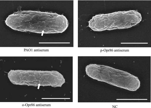
FIG. 5.
Immunogold electron micrographs of PAO1. White dots on cells (arrow) indicate gold particles. Exponentially grown cells were probed with the primary antibodies shown below each figure and developed with a secondary gold-conjugated anti-rabbit IgG antibody. NC indicates nonimmunized serum. Bar, 1 μm.

