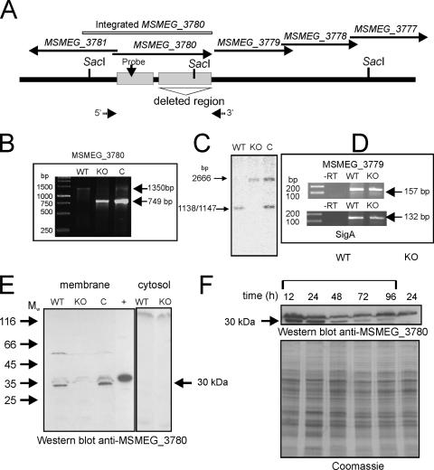
FIG. 3.
Generation and characterization of the ΔΜSMEG_3780 strain and expression of MSMEG_3780 during growth. (A) Diagrammatic representation of the genome region of MSMEG_3780 indicating the presence of SacI sites used for analysis and the region encompassing the catalytic domain of MSMEG_3780 that was deleted in the knockout strain. Arrows indicate the 5′ and 3′ primers used for PCR analysis. (B) PCR was performed using primers flanking the MSMEG_3780 gene with template genomic DNA prepared from wild-type (WT), knockout (KO), or complement (C) strains. The wild-type strain should amplify a fragment of ∼1,300 kb, while the knockout strain should amplify a fragment of ∼800 bp in length. The complement strain would show both fragments as a result of the insertion of the promoter-MSMEG_3780 fragment at the att site. (C) Southern blot analysis was performed on genomic DNA prepared from the three strains following digestion with SacI. The probe was prepared from a region containing the promoter of the MSMEG_3780 gene, which was also present in the fragment that was inserted at the att site in the complement strain. (D) Reverse transcription (RT)-PCR analysis of the MSMEG_3779 gene of RNA prepared from the wild-type and knockout strains. RNA was prepared from the two strains and reverse transcribed, and PCR was performed using primers specific to the MSMEG_3779 gene or sigA for normalization. (E) Western blot analysis was performed with cytosolic and membrane fractions prepared from wild-type, ΔΜSMEG_3780, and complemented strains (50 μg protein) using a specific antibody raised to the GST-MSMEG_3780 protein. The + indicates a lane loaded with 20 ng of purified His-tagged MSMEG_3780. Mw, weight-average molecular weight (in thousands). (F) Membrane protein was prepared from the wild-type and ΔΜSMEG_3780 strains and analyzed by Western blotting using MSMEG_3780 antibody. The Coomassie-stained gel shows the normalization of total protein taken for the Western blot analysis.

