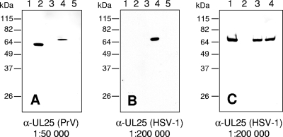
FIG. 2.
Western blot analyses. (A and B) Lysates of cells infected for 24 h with PrV-Ka (lanes 2), PrV-ΔUL25 (lanes 3), HSV-1 KOS (lanes 4), or HSV1-ΔUL25 (lanes 5) or mock-infected RK13 cells (lanes 1) were separated on SDS-10% polyacrylamide gels. After electrotransfer onto nitrocellulose membranes, parallel blots were incubated with monospecific antisera against pUL25(PrV) (A) or pUL25(HSV-1) (B). (C) Purified virions of HSV-1 KOS (lane 1), HSV1-ΔUL25 propagated on RK13-UL25(HSV-1) cells (lane 3) and PrV-ΔUL25 propagated on RK13-UL25(HSV-1) cells (lane 4) were analyzed by immunoblotting with antiserum to HSV-1 pUL25. As a control, supernatant of HSV1-ΔUL25-infected RK13 cells was processed identically (lane 2). Molecular masses of marker proteins are indicated on the left.

