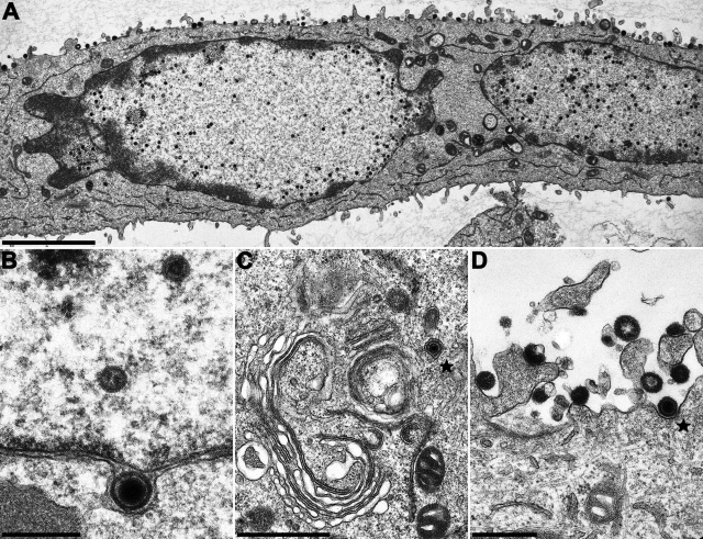
FIG. 7.
Ultrastructural analysis of PrV-ΔUL25 deleted on HSV-1 pUL25-expressing cells. RK13-UL25(HSV-1) cells were infected at an MOI of 1 with transcomplemented PrV-ΔUL25 and analyzed 14 h after infection. (A) Overview of an infected cell; (B) primary enveloped virion; (C) secondary envelopment in the cytosol; (D) extracellular L-particles and an enveloped virion. Scale bars: A, 3 μm; B, 300 nm; C, 1 μm; D, 500 nm. Virions in panels C and D are marked by asterisks.

