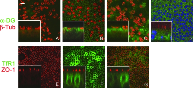
FIG. 2.
Immunolocalization of arenavirus receptors in human airway epithelia. En face (A to G) and corresponding confocal vertical sections (inset) of primary human airway epithelia following immunohistochemistry are shown. The localization of α-DG was detected by isotype control antibody (A) or α-DG IIH6 monoclonal antibody (B and C). α-DG expression is represented by a green (FITC) signal, while a red signal indicates cilium-specific β-tubulin Cy3 labeling of the apical surface. (D) TO-PRO 3 (blue) was used to label nuclei. The vertical focus in panel D lies beneath the tight junctions to display basolateral membrane labeling. Scale bar in panel A = 20 μm; n = 12 epithelia from 6 different donors. (E to G) The expression localization of TfR1 detected by secondary antibody only (E) or CD71 monoclonal antibody (F and G). TfR1 expression is represented by a green (Alexa Fluor 488) signal, while a red signal (Alexa Fluor 568) indicates tight junction boundaries detected by ZO-1 antibody. The vertical planes of focus in panels F and G differ to demonstrate unique localization; n = 10 epithelia from 5 different donors.

