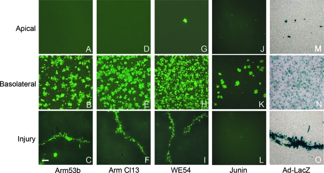
FIG. 3.
Polarity of infection by infectious arenaviruses. Well-differentiated cultures of primary human airway epithelia were analyzed to determine the polarity of virus entry following either apical or basolateral virus application. Top to bottom, the panels represent apical infection (A, D, G, J, and M), basolateral infection (B, E, H, K, and N), or apical infection following surface injury by scratching the epithelia with a pipette tip (C, F, I, L, and O). LCMV Armstrong 53b (A to C), LCMV Armstrong clone 13 (D to F), and LCMV WE54 (G to I) proteins were detected with a polyclonal antibody followed by FITC-labeled secondary antibody; n = 12 epithelia from 4 different donors. Junin protein (J to L) was detected with monoclonal antibody J3.6.2, followed by Alexa Fluor 488 secondary antibody; n = 6 epithelia from 3 different donors. As a control, Ad-LacZ (M to O) was applied and cells were stained with 5-bromo-4-chloro-3-indolyl-β-d-galactopyranoside. Scale bar (C) = 100 μm.

