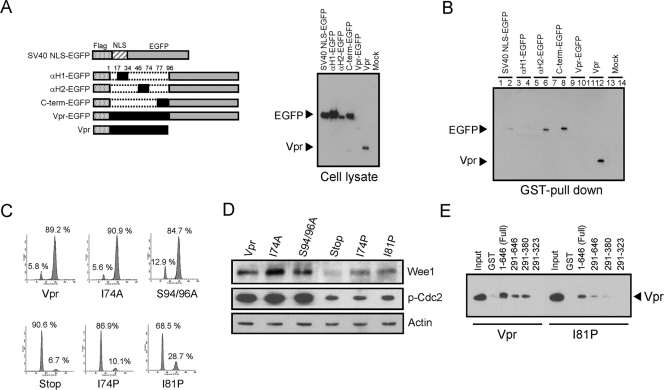
FIG. 6.
Vpr binds Wee1 through two independent domains, αH2 and C terminus domains. (A) Constructs of Flag-tagged Vpr (Vpr), αH1 (aa 17 to 34), αH2 (aa 46 to 74), C terminus (aa 77 to 96), or the NLS of the large T antigen of SV40 fused to the N terminus of EGFP. Each Vpr domain is represented in black (left panel). Above EGFP fusion proteins (EGFP) and Flag-tagged Vpr (Vpr) were expressed in HeLa cells and analyzed by Western blotting with anti-Flag antibody (right panel). (B) A total of 50 pmol each of GST (lanes 1, 3, 5, 7, 9, 11, and 13) or GST-tagged Wee1 (lanes 2, 4, 6, 8, 10, 12, and 14) preadsorbed on glutathione-Sepharose 4FF beads was incubated with 0.5 mg of HeLa cell lysates either mock transfected (mock) or transfected with expression vectors shown in panel A. The amounts of EGFP fusion proteins (EGFP) and Flag-tagged Vpr (Vpr) bound to Wee1 were analyzed by Western blotting with anti-Flag antibody. (C and D) HeLa cells were synchronized at the G1/S phase by a double-thymidine block and then infected with equivalent amounts of lentiviral vector encoding Flag-tagged wild-type Vpr or Vpr mutants as indicated. Cells were stained with propidium iodide and analyzed for cell cycle by flow cytometry. A total of 10,000 events were collected and analyzed in each sample. The number in each panel indicates the percentage of cells in each peak (C). Whole-cell lysates were separated by SDS-PAGE and probed with antibodies to Wee1, phosphorylated Cdc2 (p-Cdc2), and actin (as a loading control). There were equivalent levels of Vpr expressed from each sample (data not shown) (D). (E) A total of 50 pmol each of GST or GST-tagged full-length and deletion mutants of Wee1 preadsorbed on glutathione-Sepharose 4FF beads was incubated with 0.5 mg of HeLa cell lysate transfected with either Flag-tagged Vpr or Vpr I81P mutant (I81P) encoding expression vector. The amounts of Vpr or I81P mutant bound to Wee1 were analyzed by Western blotting with anti-Flag antibody. Input, 0.5% of cell lysates were loaded as a control for Vpr expression.

