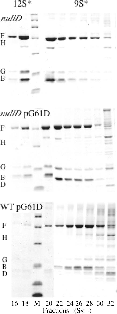
FIG. 3.
Analysis of early assembly intermediates synthesized in wild-type-infected cells. For these experiments, each gradient was prepared as described in Materials and Methods and separated into approximately 50 80-μl fractions. Every other fraction was analyzed by SDS-15% PAGE. In the figure, only the relevant fractions are depicted, i.e., fractions 16 to 30 from the bottom of the tube. The 12S* particle is located in fractions 18 to 20, and the 9S* particle is seen in fractions 26 to 28. The third lane from the left contains a molecular weight marker.

