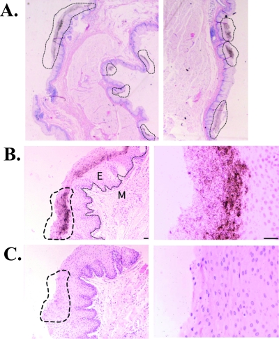
FIG. 2.
FV RNA expression localizes to the superficial epithelium. Dark silver grains overlying cells indicate an FV RNA-specific signal in the rhesus macaque pharyngeal epithelium. FV RNA was detected by ISH with a 35S-labeled FV RNA probe (679-nucleotide fragment of gag in antisense orientation) (A and B) or a control sense probe (C), using tissues that were cut into 4-μm sections and counterstained with hematoxylin and eosin. Shown are bright-field micrographs of FV RNA+ regions at ×20 (A), ×100 (left) and ×400 (right) (B and C) magnifications. Dashed lines indicate FV RNA+ regions or the same region, using the sense probe (C). A line indicates the basement membrane in panel B (left). M, mesenchyme; E, epithelium. Cells were counterstained with hematoxylin and eosin. Scale bar, 50 μm.

