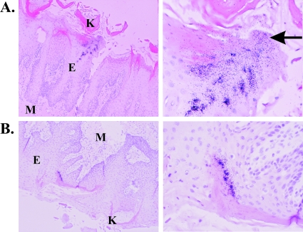
FIG. 4.
FV replication in the superficial epithelium of the tongue localizes to the superficial epithelium. Sections were treated as described in the legend to Fig. 1. Two FV+ regions from an infected tongue are shown. Cells were counterstained with hematoxylin and eosin, and the keratinized regions are demarcated by the dense pink staining. Images are shown at ×100 (left) and ×400 (right) magnifications. M, mesenchyme; E, epithelium; K, keratinized layer. A region where infected cells are sloughing from the tissue is denoted by an arrow.

